Warning signs your tongue gives you

Tongue-warnings blog

The tongue is a group of muscles that helps for tasting swallowing, speaking, etc. The surface of the tongue is covered by small bumps called papillae. Taste buds are present in papillae. The tip of the tongue is more sensitive to sweetness, both sides of the tongue to sour taste and back of the tongue to bitter taste. Salt taste sense is widely spread in the tongue. But it is greatest in the tip. The tongue is mobile and firmly attached to the floor of the mouth. The tongue is affected by oral disease and shows signs of other systemic diseases. So it is important to inspect the tongue regularly and take care of it.

Signs of disease in tongue:

Candidiases or Oral thrush!

Candidiasis is caused by a yeast-like fungus called conidia albicans. It is the most common disease in the tongue. It appears as most white patches. For infants, candidiasis is painless but for adults, causes a burning sensation. It is most common in women. Oral candidiasis is caused by poor oral hygiene maintenance and other systemic conditions like AIDS, Immunosuppression, diabetes, drugs et. Oral candidiasis is easy to wipe out with gauze. Persistent causative organism leaves ulcerative painful red lesions upon the removal of white patches. A topical anesthetic gel is given pain a burning sensation.

Leukoplakia

It is also called chronic superficial glossitis. It is caused by syphilis, smoking, sharp tooth, sepsis, spices, Leukoplakia affect anterior 2/3rd of the tongue. The affected area of the tongue shows a white patch with fissures and cracks. Treating the symptom and removing the causative is the treatment for leukoplakia.

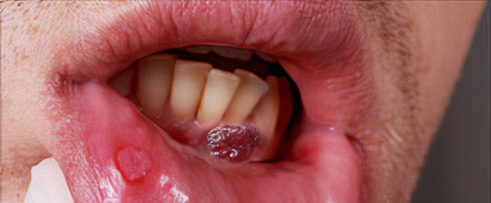
Squamous cell carcinoma:

Squamous cell carcinoma is the most common carcinoma in the tongue.  It is caused due to chronic dental trauma, tobacco smoking, candidiasis, syphilis, trauma, alcohol. The common sign of carcinoma in tongue is painless ulceration, excessive salivation, sore throat, difficult to speak, hoarseness of voice and difficult to swallow. The affected tongue is treated according to the severity.

Aphthous ulcer:

Aphthous ulcer is a painful single or multiple ulceration. This ulcer is recurrent and heals spontaneously. Aphthous ulcer is due to stress, vitamin B12, folic acid, and iron deficiency, allergy and after cessation of smoking, streptococcus sanguinis and 2 hemolytic streptococcus are causative organisms. The aphthous ulcers can be healed in 7-10 days. For treatment, vitamin supplements is prescribed and topical anesthesia to reduce pain.

Glossitis:

In iron deficiency, anemia the tongue appears smooth, glazed, fiery red, swollen, and painful tongue. It is also associated with disturbed taste and dry mouth. For treatment, iron supplements are prescribed.

Geographic Tongue:

Geographic tongue is an irregular reddish area, surrounded by white papillae. It is associated with psoriasis and lichen planus. It increases sensitivity towards spices sweet and salt. This lesion heals and recurs in other parts of the tongue. It causes pain or a burning sensation. Topical corticosteroids, topical anesthesia are given to subside symptoms. Zinc supplements and a bland diet are advised.

Black hairy tongue:

A black hairy tongue is the overgrowth of papillae of the tongue with an accumulation of keratin. It is caused by poor oral hygiene, fungal infection, bacterial overgrowth, radiation, use of drugs like penicillin, sodium peroxide. Increased length of papilla brushes and the palate cause gagging. Brushing the tongue for 2 minutes twice daily is advised. Topical agent podophyllum is prescribed.

How to clean tongue:

Tongue scraping:

Tongue scraping is used to remove debris from the surface of the tongue. The volatile sulfur compound (odor-causing substance) should be scraped off to prevent bad breath. A tongue scraper or toothbrush is used to clean the tongue. Tongue scraping is done after brushing and flossing. The scrapper is placed as far back of the tongue as possible. With gentle pressure, scrape the tongue. Rinse the scraper. Swish the mouth with water. Repeat scrapping. Another equally effective method is brushing the tongue. In this method, the toothbrush is used to clean the tongue. Gently brush the tongue for two minutes daily. These two methods remove harmful bacteria from the tongue.

Conclusion:

Every day in the morning routine while brushing teeth tongue hygiene should not be forgotten. Any changes in the tongue should never ignore. Taking care of the tongue improves taste and reduces bad breath.


dr-jovitta Dr-Jovitta

Author: Dr M. Jovitta, Pediatric /Preventive Dental Health Expert,
Jerush Dental & Facial Corrective Centre

Click to read Dr Jovitta’s other blogs: “Why flossing is important” “Brushing Techniques” “Bad breath or Halitosis” “Smoking and Oral Health”, Tooth replacement options for missing teeth” “Dental pain & Root canal Procedure”, “Coronavirus (COVID-19), Symptoms and Prevention”, “Gingivitis & Covid 19″  “Oral cancer treatment” “Pericoronitis” “Teething” “Invisalign” “Dental jewels”

சர்க்கரை நோயும் பல் இழப்பும்.

Diabetes-and-Tooth-loss

சர்க்கரை நோயும் பல் இழப்பும்

நம் வாழ்க்கை பாணி மற்றும் உணவு பழக்கம் மாறும்போது சர்க்கரை நோய் போன்ற நோய்கள் வருவதற்கான சாத்தியக்கூறுகள் அதிகமாகின்றன. சர்க்கரை நோயானது உடம்பின் நோய் எதிர்ப்புசக்தியை பாதிப்பதோடு உடலின் ஆரோக்கியத்தையே பாதிப்படையச்செய்ய  காரணமாகிறது.

வாய் மற்றும் பற்களில் ஏற்படும் பெரும்பாலான நோய்கள் மெதுவாக மற்றும் கால நீட்சியுடன் தீவிரமடைவதாலும், முற்றிய நோயை முற்றிலும் மருத்துவத்தால் குணப்படுத்துவதென்பது கடினமாக இருப்பதாலும் பல் மருத்துவரை குறைந்தபட்சம் ஆறு மாதத்திற்கொருமுறை அணுகி ஆலோசனை பெறுவதும் நோய்களுக்கு தகுந்த நேரத்தில் சிகிச்சை பெறுவதும் வழக்கமாக கொள்ளவேண்டும். இல்லையேல் பல் இழப்பு ஏற்படுவது உறுதி. அதுபோலவே நாளுக்கு நாள் அதிகமாகும் சர்க்கரை நோயாளிகளின் எண்ணிக்கைக்கு ஏற்ப பல் தொடர்பான நோய்கள் அதிகமாகிக்கொண்டே வருவதை எச்சரிக்கையுடன் கருத்தில் கொள்ளவேண்டும்.

பல் பிடுங்கும்போது சர்க்கரை நோயாளிகளின் இரத்தத்தில் சர்க்கரை அளவு கட்டுப்பாடுடன் இருக்கவேண்டுமா?

Diabetes-Checking Diabetes Checking @ Jerush

உதுதியாக ‘வேண்டும்’ என்று அழுத்தமாக சொல்லமுடியும். என்னுடைய அனுபவத்தில் நான் பார்த்த இக்கட்டான சூழல்களில் ஒன்றை குறிப்பிடுகிறேன். அருகிலுள்ள ஊரிலிருந்து ஒருவர் பல்லில் வலி இருப்பதாக சொல்லி எங்கள் பல் மருத்துவமனைக்கு வந்தார். சுமார் பத்து வருடங்களாக சர்க்கரை நோய் இருப்பதாகவும் சொன்னார். நான் அவரை பரிசோதனை செய்தபோது, பற்களுக்கும் ஈறுகளுக்கும் இடையில் அதிகமாக ‘பிளேக்’ படிமங்கள் இல்லை என்றாலும் அவரின் ஈறுகள் அதிகமாக பாதிக்கப்பட்டும் பற்கள் ஆட்டங்காணவும் செய்தது. இதிலிருந்து அவரது இரத்தத்தின் அதிக சர்க்கரை அளவு நோய் எதிர்ப்புசக்தியை எந்த அளவிற்கு பாதித்திருக்கிறது என்பது புரிந்துகொள்ள முடியும்.

நோய் எதிர்ப்பு சக்தியே எந்த நோய் பாதிப்பிற்கும்  எதிரான முன் களப்போராளி என்பதால் சர்க்கரை நோயாளிகள் தகுந்த எச்சரிக்கை உணர்வுடன் தவறாமல் பல் மருத்துவரை அணுகி ஆலோசனை பெறவேண்டும்.

தக்கலை ‘ஜெருஷ் பல் மற்றும் மூகசீரமைப்பு மருத்துவமனை’

நாங்கள் 2002 ஆம் ஆண்டிலிருந்து பல் மருத்துவ சேவையில் உள்ளோம். பல், வாய் மற்றும் முக சீரமைப்பு சிகிச்சையில் மக்கள் மனதில் சிறப்பான இடத்தை எங்கள் உலகத்தரமான சிகிச்சை வசதிகள் மற்றும் அர்பணிப்புடனான மருத்துவ சேவையின் காரணமாக பெற்றுள்ளோம். சென்னை அடையாறில் எங்கள் கிளை செயல்படுகிறது. விரைவில் நாகர்கோவிலில் புதிய கிளை துவக்கப்பட்ட உள்ளது என்பதையும் பொதுமக்களுக்கு அன்புடன் தெரிவித்துக்கொள்கிறோம்.


Author: Dr. Bladbin, MDS. (Oral & Maxillofacial Surgeon)
Jerush Dental & Facial Corrective Centre

Dental Jewels –Cosmetic Dental Treatment

dental-jewellery-coverimage

Introduction:

Nowadays people become more modernized and followers of the latest fashion. The rising generation wants to look fashionable and unique. Body tattooing, body piercing is popular now. As people are creeping up for an aesthetic look, cosmetic dentistry also upgrading cosmetic wise. People are seeking a dentist for a confident smile with an extra spark in it. Jerush Dental & Facial Corrective Centre offers all Cosmetic Dental treatments including Dental Jewels for candidates from in and around Nagercoil, Thuckalay Marthandam in Kanyakumari District, Tamilnadu and across the world

Dental Jewels:

Dental Jewellery is a cosmetic dental procedure in which diamonds, gems and other crystals are fixed on the tooth surface for a sparkling smile. Dental jewellery includes diamonds. Tooth rings, tooth tattoo, etc. The person’s tooth should be healthy to receive dental jewel.

Tooth Gems:

a-girl-wearing-dental-jewellery tooth jewellry -Girl

Tooth gems are crystals, glass mounted on a thin foil of aluminium. It is available in different colours. Skyce (Tooth Jewel) are clear, sapphire are white or blue crystals. Brilliance tooth jewellery is of three different colours, Sapphire blue, crystal clear and ruby red. They are of two sizes, 1.8mm and 2.6mm. A tooth gem is a rhinestone jewel to be attached to the tooth surface. Some dental gems are real 18k white gold and 24k or 22k gold jewellery for dental cosmetics. These jewels are made in Germany. The glass crystals have a flat back surface with a special coating. In light, these glass crystals shine like a diamond. The glass crystals are attached to the tooth surface with adhesive. Dental drill is not required for the glass crystals. Tooth gems are available in different shapes, numbers, letters and real gemstone. Glass crystals are available in 9 colours. They are diamond, rainbow, ruby, sapphire, emerald, emerald green, pink, aquamarine, sapphire light. Sapphire and Skyce are excellent dental germs. Skyce is a more shining dental gem. The dental gems are harmless to tooth as it is easy to fix and remove without any damage to the natural tooth. It does not require any tooth preparation and it is a painless procedure. Glass crystals are just like orthodontic brackets. It can be bonded to the tooth surface in the desired colour. The gem is placed with a special adhesive to avoid any damage to teeth.

How dental gems are fixed on teeth?

Firstly, to increase retention, 37% phosphoric acid is applied in enamel for 15-20 secs. The glass crystal (skyce or sapphire) is bonded to tooth with flowable transparent Heliobond. The dental gem can be fixed with flowable composite also. For bonding dental gem with composite, the tooth should be cleaned and polished with fluoride-free paste. The tooth is dried and isolated 37% phosphoric acid is applied for 15-20secs to etch the enamel surface cleaned with water and then dry the enamel for 10 secs. Apply light cure bond and light cure for 20 sec. A small amount of flowable composite is applied to the tooth and the dental gem is placed in the tooth and the dental gem is placed in the desired position. Then light cure for 40-50 sec. Now it is ready to shine like a diamond and to give an attractive sparkling smile.

Other Dental jewels:

Twinkles are pure gold and precious stones like ruby, diamonds and sapphires. They are bonded to the tooth surface. It is a painless procedure and causes no harm to natural teeth. Tooth ring is placed in the disto-incisal corner of the maxillary incisor. Perforation is prepared, polished and the ring is hung through it. While preparing tooth ring overjet should be considered, otherwise, the ring will interfere with the occlusion. Dental tattoos are designs added to the dental crown of the patient before sealed in a natural tooth. Dental tattoos include flowers, animals, patterns, even celebrities. These tattoos can be removable. Removable dental jewel, dental crown with diamonds are created according to the patient’s demand.

Complications:

  • Allergy, Aspiration, Tooth fracture.
  • Increase the chance of plaque accumulation.
  • Dental Jewellery is not recommended for high caries prone patients.
  • Metal allergy, gingival inflammation, abrasion of the tooth.

Conclusion:

Dental jewellery gives sparkling smile.  It is a safe and painless procedure. The dental jewellery trend has been gaining attraction among people of all ages. Oral hygiene maintenance and regular checkup avoid any caries development.


dr-jovitta Dr-Jovitta

Author: Dr M. Jovitta, Pediatric /Preventive Dental Health Expert,
Jerush Dental & Facial Corrective Centre

Click to read Dr Jovitta’s other blogs: “Why flossing is important” “Brushing Techniques” “Bad breath or Halitosis” “Smoking and Oral Health”, Tooth replacement options for missing teeth” “Dental pain & Root canal Procedure”, “Coronavirus (COVID-19), Symptoms and Prevention”, “Gingivitis & Covid 19″  “Oral cancer treatment” “Pericoronitis” “Teething” “Invisalign”

Invisalign – Best Orthodontic Treatment Nagercoil

invisalign-orthdontic-treatment Nagercoil

Jerush Dental & Facial Corrective Centre, The Best dental Hosptial in Kanyakumari District, provides the best orthodontic treatment including Invisalign in and around Thuckalay, Nagercoil, and Marthandam towns of Kanyakumari District.

Invisalign

Introduction:

Nowadays people are more concerned about their appearance in their professional and personal lives. As the face and the teeth have the most important role to present to the outside world adults and children are seeking dentists to improve their look. First, in early periods, malocclusions are corrected by the metallic orthodontic appliances. Then comes tooth coloured brackets and wire. So as the patients’ esthetic requirements increases, lingual brackets come up. But now Invisaligner has taken orthodontic treatment to whole another level.

Invisaligner

Invisialigner is a transparent thin rigid removable plastic aligner that helps the teeth to move in their proper position. However, this aligner meets the patient’s esthetic demand the whole crowd turn towards invisalign therapy.

invisalign-jerush-dental Invisalign, The best orthodontic treatment

How invisaligner are made?

Initially pre-treatment records are prepared that include study models,orthopantomogram and cephalometric radiographs, and clinical photos of the patient. The impression is made with polyvinyl siloxane material. Maximum intercuspation bite is recorded. The cast med was then scanned to produce a virtual 3D model. This 3D model is manipulated by the dentist to treat malocclusion with proprietary software. With this 3D model, a series of invisaligner is made.

How to use invisaligner?

The patient should wear the Invisalign for 22-24 hours every day. 10-50 invisaligners are changed every 3 week according to the movement of teeth. Each invisaligner will move the tooth to 0.25 to 0.3mm to correct the malocclusion. It takes 20-24 months for complete treatment according to malocclusion. If the patient has severe malocclusion first for few months, traditional brackets methods are used and then continued with invisaligner. Aligners should be removed while eating, drinking brushing, and flossing. Invisaligner should be cleaned with tooth brush and then soaked in 3-4 drop of chlorhexidine in lukewarm water for 5 minutes.

Who can get invisalign treatment?

Patient with mild to moderate spacing, mild to moderate crowding, orthodontically treated patient with relapse, narrow arch, deep overbite cases, distally tipped molars, mal-aligned teeth, lower incisor extraction in severe crowding. Spacing, skeletal discrepancies, short crown, multiple missing teeth, severely rotated tooth and the patient who needs premolar extraction are not the candidate for invisaligner. However initial corrections are made with the traditional bracket system and then continued with invisaligner.

Advantages of Invisaligner

  • Invisaligners are aesthetic also it gives confidence to smile and comfortable for the patient
  • Better oral hygiene maintenance
  • Short dental appointments and technically easier
  • Less allergic response
  • Removable
  • Reduce occlusal wear from parafunctional habits
  • Perfect choice for orthodontic retreatment
  • Advantage in patients with TMJ problems.
  • Low risk of enamel decalcification.
  • No root resorption.

Limitations of invisaligner

The patient should be motivated to wear invisalign for 22-24 hours. This plays an important role in the outcome of treatment. Poor oral hygiene, fracture of the aligner, skipping appointments affects the quality of the result. Invisaligner is expensive than conventional braces. In anterior open bite, skeletal discrepancies, occlusal contacts, buccolingual crown inclination occlusal relationship and overjet fixed appliance are more suggested than invisaligner.

Conclusion

The esthetic and comfort of using invisalgner plays an important role in attracting the majority of people towards invisalign treatment. The outcome of the invisalign is also promising. This leads for its success. Invisaligners are removable and easy to use. The advantages and disadvantages of invisaligner should be explained to the patient before treatment. Duration and outcome of invisalign treatment depend on patients’ cooperation. Nowadays invisaligner is more appreciated than the conventional fixed appliance.


dr-jovitta Dr-Jovitta

Author: Dr. M. Jovitta, Pediatric /Preventive Dental Health Expert,
Jerush Dental & Facial Corrective Centre

Click to read Dr. Jovitta’s other blogs: “Why flossing is important” “Brushing Techniques” “Bad breath or Halitosis” “Smoking and Oral Health”, Tooth replacement options for missing teeth” “Dental pain & Root canal Procedure”, “Coronavirus (COVID-19), Symptoms and Prevention”, “Gingivitis & Covid 19″  “Oral cancer treatment” “Pericoronitis” “Teething”

Teething

Teething

Teething is when a baby’s teeth begin to erupt in the oral cavity by breaking through the surface of gums. It may be associated with local and systemic factors that can be troublesome for babies and confusing for parents. Teething is also called odontiasis.

The eruption of primary teeth

The eruption of primary teeth (milk teeth) usually starts in the 4th – 6th month. First, two lower front teeth erupt then two upper front teeth erupt next both sides of the teeth eventually erupt. In their third year, most children have all 20 primary teeth in the oral cavity. The primary teeth (milk teeth) play an important role to preserve space for permanent teeth. During the eruption of primary teeth (i.e.) teething local irritation and other disturbance may occur.

Sign and symptoms of teething

  • The gingival (gums) around the newly erupting primary teeth will swell and become painful to touch.
  • Patches of redness on the cheeks.
  • Crying for no reason, bringing their hands toward the mouth.
  • Improper eating habits.
  • Restlessness
  • Sleeplessness and change in sleeping habits.
  • Increased salivation and drooling.
  • Increased thirst.
  • Low-grade temperature.
  • Rashes in the skin.
  • Cough, Bronchitis.
  • Diarrhea, vomiting, uncontrolled muscle contraction.
  • Bluish swelling over the erupting teeth (erupting cyst).
  • Small pieces of hard tissues over the erupting permanent molar teeth (eruption sequestrum).
  • Transmigration, transposition.

Preventive measures

  • Maintain good oral and general body hygiene.
  • A sufficient quantity of vitamins, proteins, minerals should be given.
  • Cleaning your baby’s gums should be done after each meal with cotton soaked in cold water.
  • Gentle massage to your gums to ease discomfort.
  • Keep your baby’s face clean. Always wipe out the drool to prevent rashes.
teething-child-dental-hospital-nagercoil Teething-Dental Hospital Nagercoil,
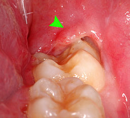
General measures:

Babies have the natural desire to chew on hard objects during teething. This helps for a painless eruption.

  • Hard unsweetened Rusk or toasted bread is given for babies to chew. It helps gingival gum) stimulation and increases blood supply to gingival. Apple, guava helps in teething and also develops taste. Keep your eyes on your baby as these foods may cause choking. Clean their oral cavities after eating, otherwise, it may lead to dental caries if primary teeth exist.
  • Pacifiers, teething keys, teething rings, teething blowers help teething. Cold pacifiers and teething toys are soothing to your baby’s gums; but not frozen as it may hurt their mouth. Clean the teething toys or pacifiers before giving them to your babies. Avoid teething products made of plastic, which may break and leads to choking. Avoid teething products made of lead which is harmful to babies. Rubber teething products, teething toys made of firm materials are recommended. Teething toys should be in one piece.
  • Teething necklaces are not recommended as they may break while chewing and have a great chance that the baby might swallow the beads.
  • Fluid-filled teething rings should be avoided because it may tear and your baby may swallow the fluid.
  • Medication that has the anesthetic effect (teething gel) should be not used as it may numb the throat and make your baby hard to swallow.
  • Medicines like Benzocaine, Ibuprofen, Aspirin causes serious side effects. Herbal and teething medications are not recommended. Some medicines that contain alcohol are toxic to babies.
  • Acetaminophen (Paracetamol) is the recommended dose according to your baby’s weight can be given to relieve pain.

When the first teeth set erupt in your baby, a soft-bristled brush and non-fluoride toothpaste are advocated.

If the baby has bleeding from gingival (gums), swelling and pus discharge from gums, high fever, vomiting, diarrhea, sleep problem, refuse to eat food, and no teeth had erupted for 2 years. Never ignore and seek medical help.

Jerush Dental & Facial Corrective Centre is a leading hospital for pedodontics. Our pediatric dentists are qualified, skilled, and have years of experience in treating child patients.


dr-jovitta Dr-Jovitta

Author: Dr. M. Jovitta, Pediatric /Preventive Dental Health Expert,
Jerush Dental & Facial Corrective Centre

Click to read Dr. Jovitta’s other blogs: “Why flossing is important” “Brushing Techniques” “Bad breath or Halitosis” “Smoking and Oral Health”, Tooth replacement options for missing teeth” “Dental pain & Root canal Procedure”, “Coronavirus (COVID-19), Symptoms and Prevention”, “Gingivitis & Covid 19″  “Oral cancer treatment” “Pericoronitis”

Pericoronitis

peri coronitis cover

Pericoronitis

Pericoronitis is an acute infection with inflammation of surrounding safe tissues of the partially erupted impacted tooth. When the third molar (Wisdom tooth) fails to erupt completely incorrect position will pave the way for pericoronitis. But it can occur around the base of any tooth that is more common in the third molar and mostly evidenced in late adolescence (20-30yrs of age).

Pericoronitis is of two types. Acute pericoronitis have severe symptoms, systemic involvement. Acute pericoronitis is seen in patients with poor oral hygiene. Chronic pericoronitis have mild symptoms and seen in patients with moderate oral hygiene.

pericoronitis-mage Pericoronitis-Photo

Causes:

Pericoronitis is caused by multiple facultative anaerobic bacteria which are peptostreptococcus, fusobacterium, and Bacteroides. When the tooth partially erupts it will be associated with soft tissue covering the tooth partially which is called the operculum. Third molars are located at the back of the mouth. So it makes it difficult to achieve effective cleaning while brushing. Impaction in this region will be high which becomes the favorite place for bacteria to grow and multiply.

Initially the overlapped tissue (operculion) inflame if not treated infection may spread to adjacent tissues and worsen the condition. Supra erupted upper third molar can cause constant trauma to the operculum and aggravate the situation. On the inner surface of the operculum, there are various degrees of ulceration.

Signs and symptoms:

  1. Pain
  2. Redness
  3. Swelling of tissue
  4. Difficulty in chewing
  5. Difficulty in opening the mouth
  6. Increase in body temperature
  7. Pain may radiate to other parts like the ear, throat, the floor of the mouth.
  8. Halitosis (bad breath)
  9. Pus discharge
  10. Bad taste in the mouth
  11. Extraoral swelling (i.e., angle of mandible)
  12. Lymphadenopathy
  13. Inability to close the jaw
  14. Loss of appetite
  15. Fatigue
  16. Leucocytosis

If pericoronitis is not treated may lead to complications. Pericoronitis convert into pericoronal abscess and may spread posteriorly into the oropharynx and to the base of the tongue leads to difficulty in swallowing. Then it causes space infection. The sequel of acute pericoronitis is peritonsillar abscess formation, cellulitis. Ludwig’s angina. It requires hospitalization and can be a life-threatening situation.

Treatment for Pericoronitis

Treatment of pericoronitis depends on the severity of the inflammation and systemic complication.

The inflamed region is irrigated with warm water or chlorhexidine solution to remove debris and exudates (pus).

The inflamed flap is deflated and cleaned with a scalar.

If pericoronitis flap is traumatized by opposing tooth, then the inflamed flap is removed or occlusal adjustment is done.

In pericoronitis with system conditions, antibiotics and analgesics are prescribed.

0.12% chlorhexidine mouthwash or warm water rinse twice a day is advised.

If the tooth is erupting in the proper position, then the inflamed pericoronil flap is removed which is called operculectomy. This procedure is done to retain the function of the tooth.

If the tooth impacted or recurrent infection occurred then extraction is the choice of treatment.

Good oral hygiene maintenance and regular follow-up is suggested.


dr-jovitta Dr-Jovitta

Author: Dr. M. Jovitta, Pediatric /Preventive Dental Health Expert,
Jerush Dental & Facial Corrective Centre

Click to read Dr. Jovitta’s other blogs: “Why flossing is important” “Brushing Techniques” “Bad breath or Halitosis” “Smoking and Oral Health”, Tooth replacement options for missing teeth” “Dental pain & Root canal Procedure”, “Coronavirus (COVID-19), Symptoms and Prevention”, “Gingivitis & Covid 19″  “Oral cancer treatment”

Oral Cancer treatment in South India

oral-cancer-cover

Oral cancer is an uncontrollable abnormal cell growth that has the potential to spread to other parts of the body. The higher the rate of oral cancer in India is attributed to the difference in lifestyle, poverty, habit, lack of education, and less access to medical care. Cancer may develop in lips, buccal mucosa (inner lining of cheeks, tongue, the floor of the mouth, hard palate. Lip cancer is more common in man and tongue cancer is more common in women. Oral cancer is life-threatening if not diagnosed and treated early. Jerush Dental & Facial Corrective Centre is a leading oral cancer treatment centre near Nagercoil & Marthandam

oral-cancer oral Cancer Treatment in South India

What increases the risk of oral cancer?

There are various etiological factors that have been involved in the causation of oral cancer.

Tobacco: The person who smokes more cigarettes daily is at more risk of developing cancer than a non-smoking person. Nicotine present in tobacco will lead to oral cancer. Chewing tobacco in the form of betel quid (paan), an areca nut, has been implicated in the causation of oral cancer.

Alcohol: The person who drinks 3 or 4 pegs of alcoholic beverage every day has twice the risk of developing oral cancer than non-drinkers. The irritants that present in alcoholic beverages damage the cells and make them vulnerable to oral cancer.

Sun exposure: Excessive sun exposure to the lip may cause lip cancer. Initially, a red erosive lesion appears then with years of sunlight exposure, the lesion becomes carcinoma.

Viruses: Oral human papillomavirus infection plays a major role in oral, head, and neck squamous cell carcinoma.

Chronic irritation: Chronic trauma from sharp teeth, ill-fitting dentures, sharp crowns, and bridges may contribute to oral cancer. The altered cell growth control processes and changes in the interaction between cells and surroundings give rise to metastasis.

Systemic factors: Vitamin A, vitamin B complex deficiency, weakened immune system, nutritional deficiency, syphilis, drugs, family history of cancer may lead to oral cancer.

Signs and symptoms of oral cancer

  1. The oral cancerous lesion present as a non-healing ulcer and bleeding from the lesion
  2. The cancerous lesion may grow exophylitically within few months
  3. The lesion has irregular edges with hardened underlying soft tissue. Then it metastasis to lymph nodes.
  4. Some cancerous lesions have pain and may have pain in the ears.
  5. Difficulty in speech, mastication, increased saliva secretion, mobile teeth.
  6. Sore throat, abnormal voice change.
  7. Weight loss.
  8. Numbness in areas of the face.
  9. Red or white patches in the mouth.

Prevention

  1. Stop smoking tobacco like cigarettes, beedis, water pipes (Hookah, Shisha, etc).
  2. Stop using smokeless tobacco like chewing betel quid (paan0, snuff, etc.
  3. Chronic alcoholics should do in moderation. It is better to stop drinking alcohol entirely.
  4. Avoid continuous exposure to the sun to your lips. Apply sunscreen lip products, wear a broad, trimmed hat to protect your skin on your lips.
  5. Take a balanced diet.

Diagnosis

Oral screening is done for patients with any irregular tissues and lumps in the oral cavity. Toluidine blue stain testing should be done to check for malignant potential. To establish the diagnosis of the lesion biopsy is done.

Treatment: The treatment of oral cancer depends on the stage, type, and location of cancer.

Surgery: Surgical removal of the lesion, cancerous lymph nodes, and metastasized other tissues are removed. The reconstruction of the excised part is replaced with dental implants and grafts to restore the function and esthetics.

Radiation: Radiation at the tumor for 2 to 8 weeks is generally given. Telecobalt machines emitting gamma rays or linear accelerators producing photons are commonly used.

Chemotherapy and targeted therapy: Drugs that kill cancer cells are given orally or intravenously. In the advanced stage of cancer, both chemotherapy and radiation therapy are given. In targeted therapy particular proteins interface their growth.

Diet: Malnutrition leads to impaired wound healing, reduced immunocompetence, and decreased tolerance to chemotherapy, radiotherapy, and surgery. It is important to get enough calories, vitamins, and minerals.

If you have any signs and symptoms that trouble you for more than weeks, make an appointment with your dentists for complete oral screening. Early diagnosis and treatment accordingly improve the chance of success.

Jerush dental & facial corrective centre, Thuckalay, Tamilnadu, South India is providing an oral cancer screening service and facility to surgically remove mouth lesions at an affordable price.


dr-jovitta Dr-Jovitta

Author: Dr. M. Jovitta, Pediatric /Preventive Dental Health Expert,
Jerush Dental & Facial Corrective Centre

Click to read Dr. Jovitta’s other blogs: “Why flossing is important” “Brushing Techniques” “Bad breath or Halitosis” “Smoking and Oral Health”, Tooth replacement options for missing teeth” “Dental pain & Root canal Procedure”, Coronavirus (COVID-19), Symptoms and Prevention, Gingivitis & Covid 19

Gingivitis – Gum treatment in Nagercoil, Marthandam

gingivitis cover

Here is a shocking revelation of research about gum disease during this COVID era! COVID-19 positive patients who are admitted in hospitals are more likely to die of breathing ailments if they suffer from periodontitis, a severe stage of gum disease. A three months study of dental researchers ended in finding the truth that Covid-19 patients with a high degree of interleukin (IL-6), a toxic protein produced by periodontitis can be a reason for life-threatening respiratory disease.

Gingivitis is a stage of gum disease prior to periodontitis. It is to be seriously taken into account by individuals about treating since it is vulnerable to periodontitis stage. Let us learn more about Gingivitis.

Gingivitis:

Gingivitis is gum disease or gingival disease. People with gingivitis often experience irritation and inflammation of gingival. Normal healthy gingival is pink in colour, maybe with some melanin pigment. The surface of the healthy gingival has an orange peel-like appearance. Proper dental care of gingivitis will easily reverse the condition of the gingiva. Taking care of gumline and paying attention to teeth will save your teeth from gingivitis. Gingivitis is one of the common presenting complaints evidenced.

gum disease-stages 
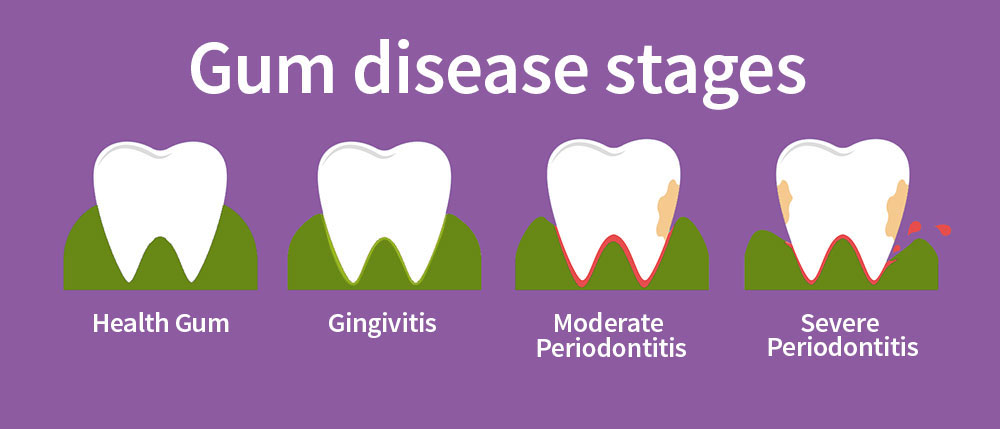
Gum Disease, different stages

How gingivitis occurs?

  1. Poor oral hygiene maintenance: Plaque is an organized bio thin film adheres to the tooth surface which mainly consists of microorganisms. If plaque is allowed to grow and mature, it causes gingivitis. The plaque hardens if it stays in your gumline which is called calculus. Plaque can be cleaned by regular brushing but for calculus, you should visit for a professional dental cleaning. The effect could get more pronounced by the accumulation of calculus because it further provides the retention of more microorganisms. If gingivitis not treated early may lead to bone loss, mobile teeth, pain, and finally tooth loss.
  2. Food impaction: Food impaction is the wedging of food between teeth. It causes radiating pain, root causes gingival inflammation and bleeding, bone loss, sensitivity, etc.
  3. Smoking: Smoking has a greater risk associated with gingivitis and can lower the success rate of treatment. People with smoking habits suffer more from gingivitis than people without smoking.
  4. Stress: Stress weakens the immunity system and negatively impacts your ability to battle against infection causes gingivitis.
  5. Poor nutrition: Malnutrition leads to an increased risk of gingivitis. Old age people with malnutrition lead to gingivitis. Vitamin deficiency weakens you and causes anemia. Gingival bleeding is the typical sign of vitamin C deficiency called scurvy.
  6. Other causes: Chewing tobacco, pregnancy, poor dental restoration, leukemia, AIDS, certain drugs, viral infections, hormonal changes, genetics, mouth breathing habit, tongue thrusting habit, bleeding disorder, toothbrush trauma, etc.

Signs and symptoms of gingivitis:

  1. Inflammation
  2. Bleeding from gingival while brushing and flossing
  3. Irritation
  4. Reddish gingival
  5. Bad breath
  6. Sensitive teeth.
Treatment:

Gingivitis resolves with good oral hygiene maintenance. Brushing twice a day for two minutes, flossing daily at least once a day will keep you off from gingivitis. When symptoms of gingivitis appear, the condition should be addressed immediately. Antibacterial mouthwash also allows, the gingiva to heal.

Professional dental cleaning and follow-up are required for a good prognosis. Damaged dental restoration should be repaired so that plaque accumulation can be prevented. Poorly fitting crown and bridges should be replaced as it may cause periodontal disease. Systemic diseases must be treated with medication. The blood sugar level should be maintained. When bone loss and other periodontal problems occur, flap surgery may be needed. Dental cleaning should be done once in six months.


dr-jovitta Dr-Jovitta

Author: Dr. M. Jovitta, Pediatric /Preventive Dental Health Expert,
Jerush Dental & Facial Corrective Centre

Click to read Dr. Jovitta’s other blogs: “Why flossing is important” “Brushing Techniques” “Bad breath or Halitosis” “Smoking and Oral Health”, Tooth replacement options for missing teeth” “Dental pain & Root canal Procedure”, Coronavirus (COVID-19), Symptoms and Prevention

Corona time. Right?

covid-right

Care during Corona Spread

Corona! by hearing this term, everyone will be frightened…upset…automatically his/her immunity will go down. Is it yes? Of course! COVID 19, Coronavirus is a group of related RNA viruses that cause disease in mammals and birds. In humans, this virus leads to RTI (Respiratory Tract Infection). Infection ranges from mild to fatal. COVID patients are increasing day by day so that getting panic does no benefit, instead, precaution helps!

Carelessness during COVID time matters. These are a few steps to be taken during this crucial time to prevent this type of infection until vaccination is discovered and make it practically applicable.

Preventive measures for Covid-19

Remember:

  1. Wash your hand with soap and water for at least 20 seconds (This period of time is important for the virus to destroy).
  2. Use of Alcohol-based sanitizer. While you come out of your house, like public places, when difficult in use of handwashing with soap, use an alcohol-based sanitizer (minimum 60% alcohol). But beware to wash your hands with soap before eating food. Don’t use a hand sanitizer nearby a stove, as it can inflame.
  3. Avoid touching your eyes, nose, and mouth without proper cleaning of hands as it can cause entry of Covid-19 virus to the body and thereby infection.
  4. Avoid junk foods. Eat healthy foods that provide adequate nutrition. Take vitamins more especially C and D during this COVID time to boost your immunity. Our body’s immune system is more important to fight against infection.
  5. Use of face masks. Proper use of face masks will prevent infection. Use it and don’t throw it to public places as it is already contaminated. Masks once it is used on your face, don’t use it next time to the opposite side, as the contaminated surface meets your face. Always remove your masks with an elastic cord.
  6. N-95 mask- best mask for more protection.
  7. Stay at home unless it is absolutely necessary.
  8. Give more importance to personal hygiene.
  9. Avoid drinking cool drinks. Drink hot water occasionally.
  10. Keep social distancing.
n95-mask 1. How to remove facemask. | 2. N95-Mask

“Prevention is better than Cure”     

Here at Jerush Dental & Facial Corrective Center, a leading Dental Hospital at Thuckalay, we maintain all new set-up and procedures as per the Government norms for a Complete Covid-Free environment for the safety of our patients and the staff. ‘Jerush’ Dental Clinic is located at a place easily accessible since it is very nearer to Colechal (colachel), Karungal, Eraniel, Monday market, Azhagiamandapam, Mulagumoodu, Marthandam, Palliyadi, Villikuri, Manali, Kumarakoil, Parvathipuram, Chunkankadai, Mandaicaud, Kulasekharam, etc. places.


dr-aiswarya Dr. Aiswarya

Author: Dr. Aiswarya BDS (Reg. No. 20083), A skilled dentist who had passed out with flying colours in her professional studies, serving more than five years at Jerush Dental & Facial Corrective centre. Other blogs: Dark Chocolate, Oral Health Advice

Dental Hospital Nagercoil Marthandam

covid19-cover

Jerush Dental & Facial Corrective Centre is the best Dental Clinic  / Dental Hospital and Covid-19 norm strictly following hospital near Nagercoil and Marthandam. Just a twenty minutes travel from Nagarcoil or Marthandam will do to reach this Centre at Thuckalay. Started as a small clinic in 2002, now they have grown to provide world-class dental care with state-of-the-art technology equipment. Karungal, Colachel, Monday Market, Kulasekharam, Azhagiamandapam are some of the places close to Jerush dental & facial corrective centre, Thuckalay, Tamilnadu. Here, all our Dentists are well qualified and skilled. Our talented Dental surgeons are looking forward to serving your dental needs with the utmost care and maximum satisfaction.

About Coronavirus

Coronavirus is a tiny group of RNA viruses, achieved a heavy impact on several major countries in the world.

How coronavirus spread?

Coronavirus spread through droplets from the nose or from the mouth as a result of coughing or sneezing of a coronavirus infected person. Coronavirus can be transmitted directly if the infected person is in close contact with a healthy person when the infected person cough or sneezes the droplets expelled have very high contagious to the person who is within one meter. The droplets which contain virus can be transmitted indirectly as it survives on clothes or other surfaces. Coronavirus survives over metal and glass for 5 days, over wood for 4 days, over plastics, and stainless steel for 2-3 days.  When the person touches any of the infected clothes and then touches his mouth, nose, or eyes will be infected by coronavirus.

What are the symptoms of COVID-19?

Symptoms of COVID-19 is very much similar to common flu. Common symptoms of COVID-19 are shortness of breath, fever with the gradual increase in temperature, cough, fatigue, chills, headache, sore throat, muscle aches, loss of taste or smell, confusion, excessive drowsiness, persistent pain in the chest, blue face or lips, diarrhea, conjunctivitis, rashes on the skin, discoloration of finger or toes, loss of speech or movement. Generally, it takes 2-14 days to show up the symptoms of COVID-19. People at any age can be infected by coronavirus. But most often, elderly persons and persons with pre-existing medical conditions like diabetes heart disease, asthma et., are more affected by coronavirus. Smoking increases the risk to be infected by coronavirus. Some researches show men are more vulnerable than women by coronavirus.

Protective and preventive measures against coronavirus:

covid-19-prevention Coronavirus Prevention
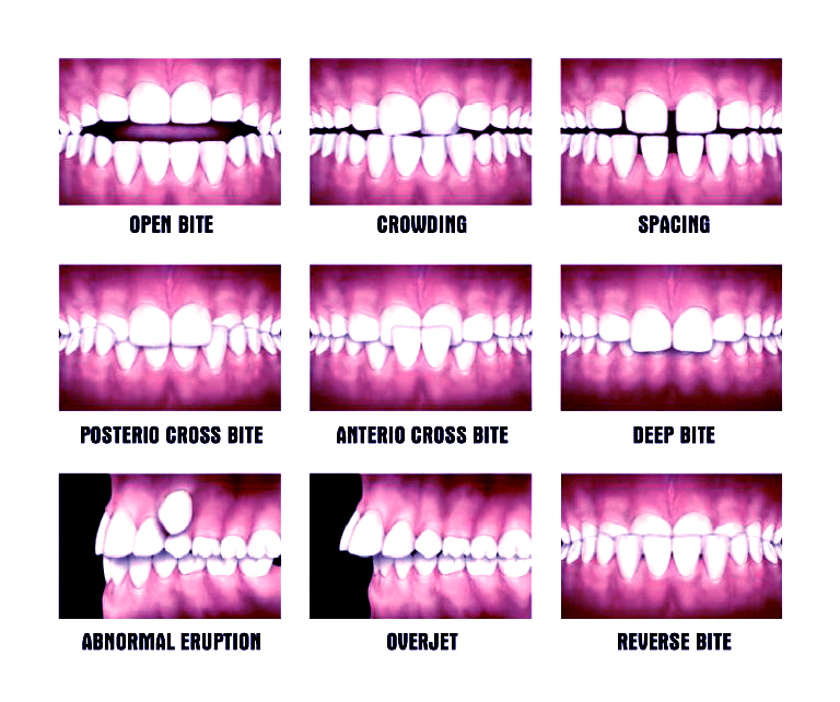
  1. Washing your hands with soap and water for 20 sec. will be effective against coronavirus. If soap and water is not available, use alcohol-based sanitizer with at least 60% ethanol or 70% Isopropanol. Hands should be washed after handling money, after using public transport, after touching the elevator button or door handles, after using ATMs, after shopping, etc. Avoid touching nose mouth eyes.
  2. Wear a mask, when you are in public places. Wash your hands before touching the mask, check the mask, for any dirt or damage before wearing masks. Hold the face mask by ear loop. Place it around your ears, secure the mask as it covers your nose, mouth, and chin. Wash your hands before removing the face mask, only touch both the ear loops and gently lift and remove the face mask. Then wash your hands with soap. This is the recommended way to use a face mask.
  3. Avoid going to places that were crowded as it causes a serious spread of coronavirus.
  4. Keep at least a 1-meter distance between yourself and other people.
  5. It is wise to stay home and isolate yourself even with minor symptoms.
  6. If you have difficulty in breathing, fever, cough, seek medical help.

Protect yourself by following preventive measures and let us together fight against Coronavirus pandemic.


dr-jovitta Dr-Jovitta

Author: Dr. M. Jovitta, Pediatric /Preventive Dental Health Expert,
Jerush Dental & Facial Corrective Centre

Click to read Dr. Jovitta’s other blogs: “Why flossing is important” “Brushing Techniques” “Bad breath or Halitosis” “Smoking and Oral Health”, Tooth replacement options for missing teeth” “Dental pain & Root canal Procedure”

Braces / Clips treatment in Nagercoil

malocclusion

Braces / Clips treatment for Malocclusions

Dental malocclusion means the abnormal alignment of the upper and lower teeth.

Etiology: Childhood habits such as thumb sucking, tongue thrusting, using pacifiers beyond age 3, and prolonged use of bottle feeding, extra teeth, lost teeth, impacted teeth, or abnormally shaped teeth.

Different types of malocclusions:

  1. Overjet
  2. Overbite
  3. Anterior crossbite (underbite)
  4. Spacing
  5. Diastema
  6. Missing teeth
  7. Impacted tooth

Classification of Malocclusion:

malocclusion-types Malocclusion-types

In 1894, Edward H. Angle published the first classification of the malocclusion.

Class I Malocclusion: Neutrocclusion: It is the most common malocclusion with normal bite and light overlap of teeth.

Molar relation: The Mesiobuccal cusp of the upper first permanent molar occludes with the mesiobuccal groove of the lower first molar. But the line of the occlusion is altered in both the arches because of individual tooth irregularities [ Crowding/spacing/rotations of other localized problems of tooth and inter-arch problems (open bite/deep bite/crossbite)]

Class II Malocclusion: (Distocclusion/Retrognathism,/overbite ) This occurs when upper jaw and teeth severely overlap the lower jaw and teeth.

Molar relation: The mesiobuccal cusp of the maxillary first permanent molar occlude mesially to the mesiobuccal groove of the mandibular first molar.

Class II Division I

Class II Division II

Upper incisors are proclaimed Upper central incisors show lingual inclination and maybe overlapped by upper lateral incisors
Excessive overjet and deep overbite Deep overbite
V-shaped upper arch Broad upper arch
Failure in anterior lip closure Normal lip seal. Deep mental groove.

Class III Malocclusion: Mesiocclusion/Prognathism,/Anterior crossbite,/negative overjet/underbite). This occurs when the lower jaw protrudes forward causing the lower jaw and teeth to overlap the upper jaw and teeth.

Mollar Relation: The mesiobuccal cusp of the maxillary first permanent molar occludes distally to the mesiobuccal groove of the mandibular first molar.

Treatment for Dental Malocclusion:

Management of Class I Malocclusion: Spacing and crowding can be corrected by fixed orthodontic braces. This treatment requires 6 months to 1 year, depending on the patient’s bone density.

Crowding can be corrected by placing braces to correct the position of the teeth by the removal of teeth to correct overcrowding, surgery to reshape or shortening the jaw wires or plates to stabilize the jaw bone.

Management of Class II malocclusion:

  1. Orthodontic Braces | 2. Conventional Orthodontics | 3. Surgical intervention | 4. People with retrognathic mandible may also develop this disorder

Management of Class III Malocclusion: The most common treatment of prognathism is the combination of orthodontics and orthognathic surgery. Dental prognathism can be treated by fixing orthodontic braces (camouflaged orthodontic treatment) mouthguard.

This fixed orthodontic treatment requires 1 year for completion. After completion of fixed ortho treatment, retainers should be placed for bone stabilization.

Jerush dental & facial Corrective Center, Thuckalay has located just 20 minutes away from Nagercoil, Marthandam, Kaliyakkavilai, Kulasekaharam, Colachel, Karungal, Monday market, Eraniel, Prvathipuram, etc.


dr-sangari Dr. Sangari

Author: Dr. Sangari, One of the Top Dentists in Jerush Dental & Facial Corrective Centre with more than a decade of experience in dental patient care. Very patient friendly and anyone would feel like a neighborhood doctor. Dr. Sangari’s long term dedicated association in our center, proved like an inevitable member among team surgeons, in creating millions of happy smiles.

Teeth whitening in Nagercoil & Marthandam

teeth-whitening

About Teeth Whitening

It is the most popular aesthetic dental treatment. Nowadays people are more interested in whitening their teeth for an attractive smile. Jerush Dental & Facial Corrective Centre is one of the leading TEETH WHITENING centers near Nagercoil and Marthandam.

Dental stains:

The cause of dental stains should be examined for better results. There are two types of stains.

dental-sains 
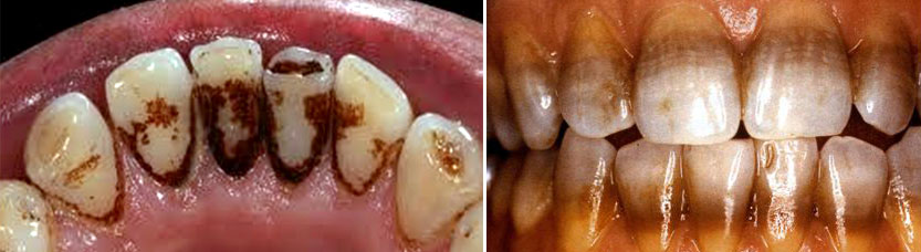
Types of dental stains 1. Intrinsic Stain 2. Extrinsic Stain

1. Extrinsic stain
2. Intrinsic Stain

Extrinsic stain means the accumulation of chromatogenic substance on the tooth surface. This type of strain occurs due to tobacco use, poor oral hygiene, ingestion of chromatogenic food, and drinks. With time this type of stain becomes darker and persistent. It can be removed by oral prophylaxis and bleaching if required.

Intrinsic stain:

An intrinsic stain is a stain that occurs interior of the teeth. This type of stains caused by aging, enamel micro-cracks, tetracycline medication, excessive fluoride ingestion, severe jaundice in infancy, dental caries, thinning of the enamel layer, traumatized tooth, etc. Intrinsic stains cannot be removed by oral prophylaxis. Only bleaching of teeth is required for a good result.

Vital tooth bleach:

In-office bleaching:

This procedure is done under the complete control of the dentist. When the desired shade achieved, the procedure is stopped. High concentrated teeth whitening agent is used. The wanted result can be achieved in a single sitting, but more sitting may be needed for better results. A 35% concentration of hydrogen peroxide is used for bleaching.

At-home bleaching:

This procedure involves a dentist-supervised night guard and carried out by patients themselves. It is self-administrated by the patient. Low cost, fewer side effects, and safer. It is a commonly practiced technique as whitening the teeth at home is possible. Bleaching gel is applied in the teeth along with a custom-fabricated mouthguard is worn at night for at least 2 weeks.

The patient should be instructed to brush his/her teeth prior to the application of the tray. The patient often forgets to do this procedure and is technically sensitive. Accurate concentration should be used as recommended by the dentist, otherwise, it may cause teeth sensitivity, gingival irritation, nausea, and throat irritation. 10% to 20% of carbamide peroxide is used.

Over-the-counter bleaching products are increased in recent days. These products are self-applied and widely used. It includes low concentrated whitening agents, whitening strips, whitening dentifrices, etc. It is recommended to use two weeks twice per day.

Non-vital tooth bleach:

In this technique, whitening agents are sealed in the pulp chamber and activated by heat or light. This procedure is repeated until the desired result is achieved.
Macro abrasion is used for the removal of localized surfaced stains. Composite finishing bur is used to remove the surface defect. The polishing is done.

Porcelain veneer:

Hollywood smile makeover is one of the popular teeth aesthetic treatments. In this procedure, teeth are corrected in perfect shape, size and a porcelain veneer is placed over the teeth. This gives perfectly aligned attractive teeth.

These are some teeth whitening techniques practiced widely. According to the type of stain, type of defect, teeth whitening technique may differ. Oral hygiene maintenance is the key to preserve the result.

Jerush Dental & Facial Corrective Centre Thuckalay is one of the top Teeth Whitening centers in Nagercoil in the Kanyakumari District. People from Marthandam, Colachel, Karungal, Monday Market, Parvathipuram, Kulasekharam, Kuzhithurai, Melpuram, and other places in the district also visit our center for professional Teeth whitening, Dental Bleaching, Dental Cleaning, etc. Our qualified and skilled lady surgeons deserve all the praise for the successful flow of dental whitening candidates, especially from Nagercoil and Marthandam in Tamilnadu.


dr-jovitta Dr-Jovitta

Author: Dr. M. Jovitta, Pediatric /Preventive Dental Health Expert,
Jerush Dental & Facial Corrective Centre

Click to read Dr. Jovitta’s other blogs: “Why flossing is important” “Brushing Techniques” “Bad breath or Halitosis” “Smoking and Oral Health”, Tooth replacement options for missing teeth” “Dental pain & Root Canal Procedure”