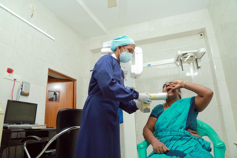
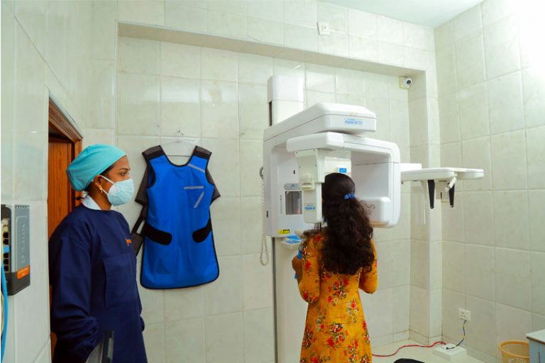
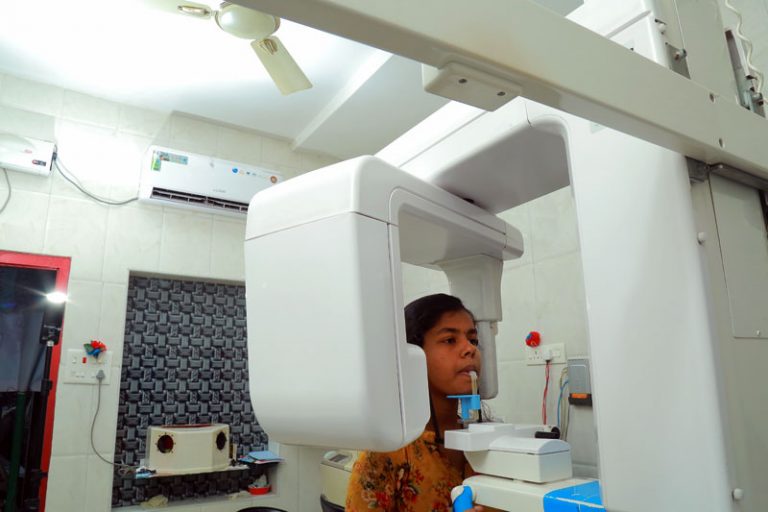
cbct scan, OPG &
x-rays


imaging options
Orthopantamogram ♦
Cephalo Lateral View ♦
Cephalo Frontal View ♦
Towen’s View ♦
♦ Water’s View
♦ TMJ Frontal View
♦ Maxillary Sinus View
♦ Others (Specify)
what are orthopantomogram & cephelogram
cbct: advantages
♦ Allows the 3D visualization of the craniofacial skeleton in patients with cleft lip and palate
♦ Ascertains tooth position and localization
♦ Useful in knowing bone dimensions and investigating the site for implant placement
♦ Helps in the treatment to correct maxillary transverse deficiency problems in adolescents.
♦ Helps to find complex conditions such as obstructive sleep apnea (OSA) and enlarged adenoids
♦ Supports in age assessment and Investigation of orthodontic-associated paraesthesia
♦ Supports in dental caries diagnosis, Periodontal and periapical disease assessment,
♦ Helping the endodontists in many ways. Enables the visualization of the anatomic complexity, disturbances in the typical morphology, presence of material in the root canal, progression, regression and maintenance of apical periodontitis.
♦ Useful in successful surgical planning of impacted teeth
♦ Useful in analyzing unusual bony lesion. cysts, tumours and a wide range of esoteric lesions…
♦ Useful in detecting trauma to teeth and alveolar bone, supports orthognathic surgery, Temporomandibular joint (TMT) surgery etc. etc…..
intraoral x-ray
The most commonly taken dental X-rays are Intraoral X-rays. These X-rays assist the dentists by providing plenty of information on cavities, health of the teeth and jaw bone, monitoring the development of new teeth. Also assists the patients to understand the real condition of their teeth before any treatment.
Types of Intraoral X-Rays
There are different types of dental X-rays available that depend on its image rendering features. Very common are
- Bitewing X-rays
- Periapical X-rays
- Occlusal X-rays
Each X-ray shows the entire arch of teeth in either the upper or lower jaw.


